Three-dimensional (3D) printing technology has emerged as one of the most important tools in the ever-evolving space of tissue engineering. But 3D printing living tissues, referred to as bioprinting, presents significant challenges, which a team in the Department of Biomedical Engineering at Texas A&M University is working to address.
One challenge in bioprinting is the need for the bioink, or the material being printed, to solidify quickly after printing to hold the desired shape. At the same time, ideal bioinks should also be able to print under low shear force so as to not damage living cells that are dispersed within the material.
“This requires very specific properties that are not easy to achieve with traditional bioinks,” said Dr. Daniel Alge, one of the researchers on the project and an associate professor in the biomedical engineering department. “Hydrogel microparticle (HMP) bioinks make it easier and significantly expand the toolkit of soft biomaterials that we can use in 3D bioprinting.”
HMPs can be used to print soft tissue-like structures because the microparticles can move past one another when pressure is applied, but are also sticky enough to hold a 3D shape after being printed. As an emerging bioink, there are still unanswered questions about printing with HMPs, since they behave differently from other bioinks. That is what the team researched and recently published in a paper about in the journal Science Advances.
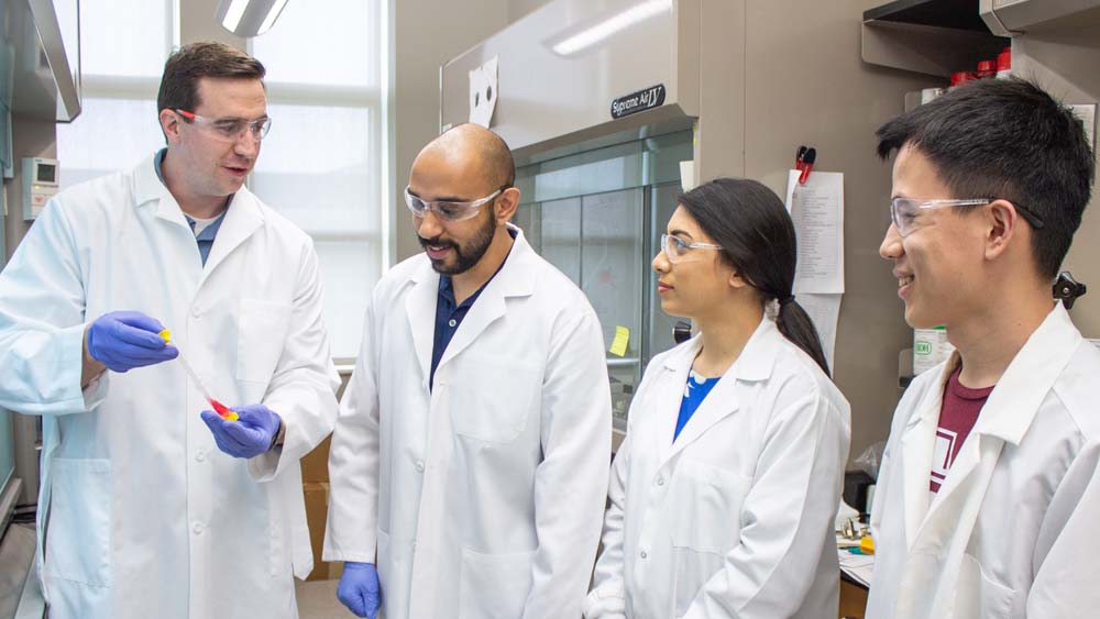
The project includes several biomedical engineering faculty: Alge, Dr. Akhilesh Gaharwar, associate professor, and Dr. Abhishek Jain, assistant professor.
In their study, the team sought to understand factors that affect how HMPs are extruded from a syringe and nozzle during printing. They specifically studied how the particles jam and then flow. Through a series of experimental and computational methods, the team found the interplay between external resistance from the printing apparatus and the physical properties of the microparticles. They also showed there is a tradeoff between factors that affect print fidelity and the viability of cells contained within the bioink.
“Bioprinting is an exciting field with enormous potential for meeting the need for transplantable organs and tissues,” Alge said. “However, in order to advance bioprinting from the realm of science fiction to reality, we need innovative materials like HMP bioinks. Ultimately, our findings can be used to improve the printability of HMPs and facilitate their broader use in 3D bioprinting.”
The team aims to apply their knowledge about how to engineer and bioprint with HMP bioinks to the production of functional tissues. Their target is a bio-artificial pancreas to treat diabetes, a project that the team received an X-Grant from the Texas A&M President's Excellence Fund to pursue. In Type 1 diabetes, the body does not produce insulin because pancreatic beta cells are attacked by the immune system. The goal is to design a 3D-printed bio-artificial pancreas with a vascularized network to protect encapsulated islets that contain the beta cells.